Recent POST


When your lungs can’t expand fully, even simple tasks like walking to the mailbox or tying your shoes become exhausting. That’s often the reality for people with pleural effusion-a buildup of fluid between the layers of tissue lining the lungs and chest wall. It’s not a disease on its own, but a sign something deeper is wrong. And if left unaddressed, it can lead to serious complications-or even shorten your life.
What Causes Pleural Effusion?
Pleural effusion happens when fluid leaks into the space around your lungs. But why? The answer depends on the type of fluid. Doctors divide it into two main categories: transudative and exudative.Transudative effusions are caused by pressure imbalances in your body. Think of it like water seeping through a weak spot in a dam. The most common cause? Congestive heart failure. It accounts for about 90% of these cases. When your heart can’t pump blood efficiently, pressure builds up in your veins, pushing fluid out into the pleural space. Other causes include liver cirrhosis-where your liver stops making enough protein-and nephrotic syndrome, where your kidneys leak too much protein into your urine. These conditions don’t involve inflammation. They’re about physics, not infection.
Exudative effusions are more serious. They’re caused by inflammation, infection, or cancer. Pneumonia is the number one culprit, responsible for 40-50% of cases. When bacteria invade your lungs, your immune system responds by flooding the area with fluid and white blood cells. Malignancy comes next-cancer, especially lung or breast cancer, spreads to the pleura and triggers fluid production. About 30-40% of exudative cases are cancer-related. Pulmonary embolism, tuberculosis, and autoimmune diseases like rheumatoid arthritis also contribute.
Here’s the key: you can’t treat pleural effusion without knowing its cause. A 2020 American Thoracic Society guideline says every effusion larger than 10mm on ultrasound must be analyzed. Why? Because 25% of cases initially labeled "undetermined" turn out to be cancer. Missing that means missing your best chance to survive.
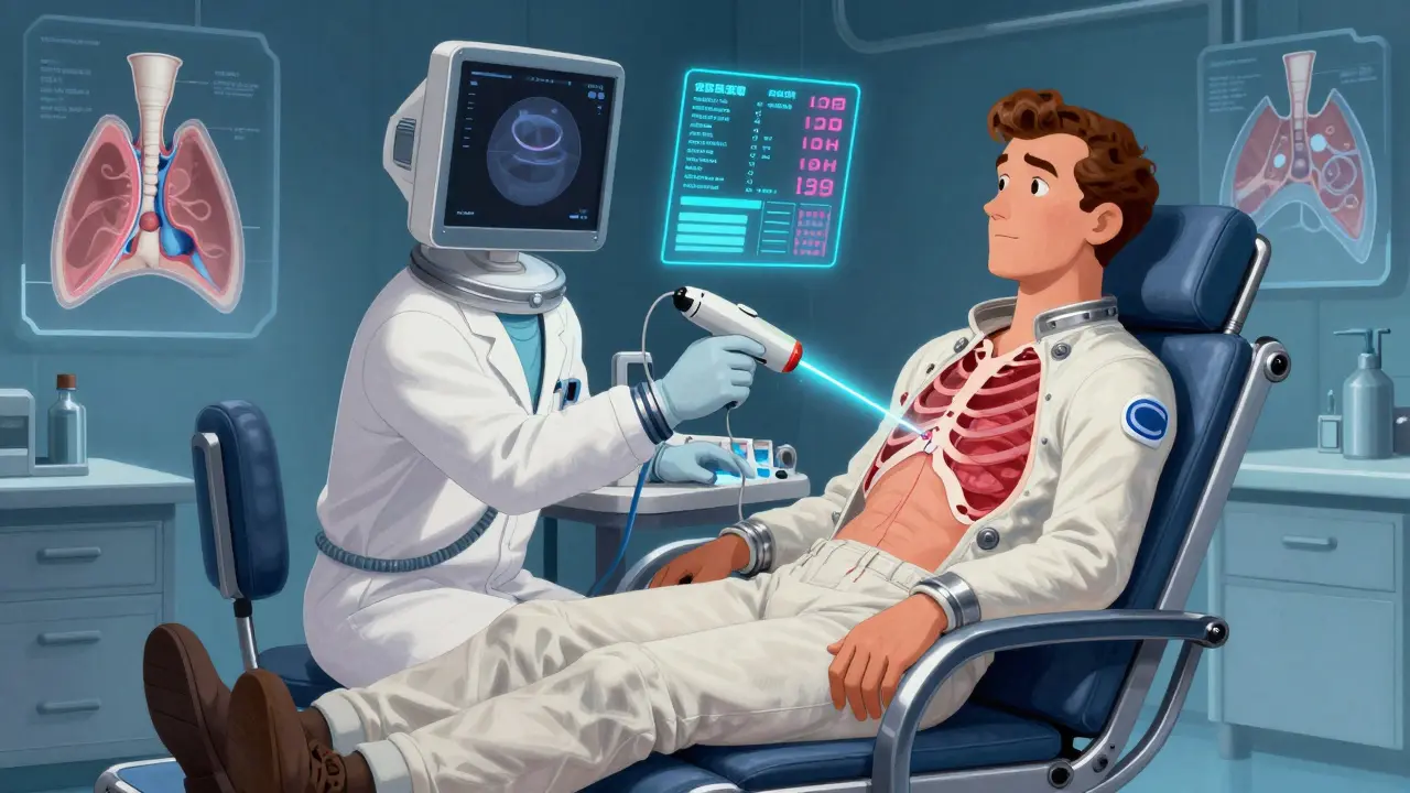
How Thoracentesis Works-and Why Ultrasound Is Non-Negotiable
If you’re short of breath and imaging shows fluid, your doctor will likely recommend thoracentesis. It’s a simple procedure: a thin needle or catheter is inserted into your chest to drain the fluid. But it’s not as simple as it sounds.For decades, doctors did this "blind," relying on landmarks like rib spaces. That changed after studies showed complication rates as high as 18.9%. Now, ultrasound guidance is the standard. It cuts the risk of puncturing your lung (pneumothorax) by 78%. That’s not a small improvement-it’s life-changing.
The procedure usually happens at the 5th to 7th intercostal space along the mid-axillary line. You sit upright, leaning forward slightly. The area is numbed. Then, under real-time ultrasound, the needle goes in. For diagnosis, they take 50-100 mL. For relief, they can remove up to 1,500 mL in one session. But here’s the catch: removing too much too fast can cause re-expansion pulmonary edema-a rare but dangerous condition where your lung swells as it re-inflates. That’s why doctors now use pleural manometry to monitor pressure. Keeping it under 15 cm H₂O reduces this risk dramatically.
The fluid gets tested for protein, lactate dehydrogenase (LDH), cell count, pH, glucose, and cytology. These numbers tell the story. A fluid-to-serum protein ratio over 0.5? That’s Light’s criteria-confirming an exudate. A pH below 7.20? That’s a red flag for complicated pneumonia. Glucose under 60 mg/dL? Could be empyema or rheumatoid arthritis. LDH over 1,000 IU/L? Often cancer. Cytology finds cancer cells in about 60% of malignant cases. But even if it’s negative, you can’t rule out cancer. Sometimes you need multiple samples or a biopsy.
Why Recurrence Is Common-And How to Stop It
Draining fluid gives you relief. But if you don’t fix the root cause, it comes back. Fast.For malignant effusions, recurrence within 30 days after thoracentesis alone is around 50%. That’s why doctors don’t stop at drainage. The goal is to seal the space so fluid can’t build up again. The two main options are pleurodesis and indwelling pleural catheters.
Pleurodesis means sticking the lung to the chest wall. Talc is the gold standard-70-90% success rate. It’s a powder injected through a chest tube. The irritation causes inflammation and scarring, which fuses the layers. But it’s painful. Up to 80% of patients report moderate to severe pain after the procedure. Hospital stays average 5-7 days. And it doesn’t work if your lung is trapped-meaning it’s stuck by scar tissue and can’t expand.
Indwelling pleural catheters (IPCs) are changing the game. These are thin tubes left in place for weeks. You or a caregiver can drain fluid at home, a few ounces at a time. Success rates? 85-90% after six months. Hospital stays drop from 7.2 days to just 2.1 days. Patients report better quality of life. And it works even when the lung is trapped. For many with advanced cancer, it’s the best option-not because it cures, but because it gives control.
For heart failure-related effusions, the fix isn’t surgery. It’s medication. Diuretics like furosemide, plus ACE inhibitors and beta-blockers, can reduce recurrence to under 15% in three months. Monitoring NT-pro-BNP levels helps doctors adjust doses before symptoms return. No need for repeated thoracentesis.
Parapneumonic effusions-those from pneumonia-need antibiotics and drainage if they’re complicated. Criteria? pH under 7.2, glucose under 40 mg/dL, or positive Gram stain. If you skip drainage, 30-40% of these turn into empyema: pus in the chest. That requires surgery.
Post-surgical effusions, especially after heart bypass, often go away on their own. But if you’re draining more than 500 mL per day for three days straight, you need a chest tube. Left untreated, they can lead to long-term breathing problems.

The Big Picture: Treat the Cause, Not Just the Fluid
Dr. Richard Light, who created the diagnostic criteria still used today, put it perfectly: "Treating the effusion without treating the cause is like bailing water from a sinking boat without patching the hole."That’s why modern medicine has shifted from symptom management to root-cause resolution. You don’t just drain fluid-you analyze it, classify it, and match it to the right treatment. For cancer, that means IPCs. For heart failure, it’s optimized meds. For infection, it’s antibiotics and timely drainage.
And the data backs this up. Between 2010 and 2020, five-year survival for patients with malignant pleural effusion jumped from 10% to 25%. Why? Better cancer therapies. Earlier diagnosis. Personalized treatment. It’s not magic. It’s precision.
Still, mistakes happen. A 2019 JAMA study found that 30% of thoracenteses were done on small, asymptomatic effusions-and provided zero benefit. That’s unnecessary risk. Ultrasound helps here too: if the fluid is less than 10mm and you’re not short of breath, watch and wait.
What You Can Do
If you’ve been diagnosed with pleural effusion:- Ask for ultrasound-guided thoracentesis. Don’t accept a blind procedure.
- Insist on full fluid analysis-protein, LDH, pH, glucose, cytology. No shortcuts.
- Know your cause. Is it heart failure? Cancer? Infection? Your treatment depends on it.
- If it’s cancer and you’re not a candidate for surgery, ask about indwelling pleural catheters. They’re not a last resort-they’re a better option.
- If it’s heart-related, stick to your meds. Don’t skip doses. Monitor your weight daily. Sudden weight gain means fluid is returning.
- Ask about pleural manometry during drainage. It’s not widely available everywhere, but it reduces complications.
Pleural effusion isn’t a death sentence. But it’s a signal. Listen to it. Act on it. And make sure your care team treats the cause-not just the symptom.

Travis Craw
man i had no idea thoracentesis used to be done blind. my uncle went through this last year and they used ultrasound the whole time - he said it was weird seeing his own lung on the screen but also kinda reassuring. guess i’m lucky they didn’t just poke around like it’s a game of darts.

Riya Katyal
oh so now we’re supposed to believe talc is the "gold standard"? sure. next they’ll tell you putting glitter in your chest cavity is "evidence-based". i’ve seen people cry for days after pleurodesis. who’s really benefitting here - the patient or the hospital billing department?

swarnima singh
you know what’s really sad? they talk about "precision medicine" like it’s some miracle, but half the people in this country can’t even get a basic ultrasound. i work at a clinic where they still use stethoscopes to guess if there’s fluid. and then they wonder why people die. it’s not the disease. it’s the system. we’re not treating patients. we’re treating insurance forms.
and don’t even get me started on how they charge $12k for a catheter that costs $40 to make. capitalism doesn’t care if you can breathe. it just cares if you can pay.
my cousin had an IPC. she drained it herself every other day. said it felt like having a little window into her own body. that’s the kind of dignity they never mention in the journals.
they say "treat the cause" - but what if the cause is poverty? what if the cause is being a single mom who works two jobs and can’t afford the follow-up? does that count as a "cause" in their algorithm?
and why is it always the poor who get the painful procedures and the rich who get the comfy catheters? it’s not medicine. it’s a class war wrapped in a white coat.
i’m not saying don’t drain the fluid. i’m saying don’t pretend this is fair.
they want us to trust science? then fix the system first. or stop pretending you care.

Isabella Reid
really appreciate how thorough this is - especially the part about pleural manometry. i’ve never heard of that before. my mom had a malignant effusion last year and they just drained her until she felt better. no pressure monitoring, no follow-up plan. she ended up back in the ER two weeks later. this article feels like a lifeline for people who’ve been gaslit by the system.
also, the IPC point? huge. my aunt had one and she said it gave her back her life. she could travel, cook, even go to her granddaughter’s recital without fearing she’d suffocate. it’s not about curing cancer - it’s about letting people live while they’re still here.
thank you for writing this. i’m sharing it with every person i know who’s dealing with this.

Bianca Leonhardt
you people are so naive. this whole thing is a scam. they make you think you’re getting care, but they’re just milking you for cash. ultrasound? sure. but did you know the machine costs $200k and they charge $500 per scan? they don’t care if you live or die - they care about your deductible. and don’t even get me started on how they push talc because it’s cheaper than surgery. it’s not medicine. it’s a profit center.
and the "5-year survival jumped to 25%"? that’s because they started diagnosing earlier - not because the treatment got better. they just caught more people before they died. that’s not progress. that’s statistics magic.
they want you to trust the system. don’t. trust your gut. if they’re pushing a procedure, ask why. always ask why.

Jody Fahrenkrug
my dad had a parapneumonic effusion after pneumonia. they drained it, gave him antibiotics, and sent him home. no pH test, no glucose check. he got worse. turned into empyema. had to have surgery. they didn’t even test the fluid properly. this article should be mandatory reading for every ER doc.

kanchan tiwari
they’re hiding something. why is no one talking about the fact that talc contains asbestos? i did my own research - it’s in the FDA’s warning database. they’re poisoning people under the guise of "treatment". and the catheters? they’re implanted with trackers. the government’s monitoring our lung fluid. you think that’s a coincidence? think again.
the real cause of pleural effusion? 5G towers. and the fluid? it’s not water. it’s nanobots. they’re in your blood. they’re in your lungs. they’re in your dreams. they want you to believe this is just pneumonia. it’s not. it’s a takeover.
ask your doctor if they’ve ever seen a patient with blue snot. if they say no - they’re lying.

Bobbi-Marie Nova
okay but can we talk about how wild it is that a 10mm fluid line on ultrasound means you need to test it? that’s like the size of a grape. i had a chest x-ray last year and they said "nothing to worry about" - turns out i had 8mm. no one said a word. if this guideline had been around then, maybe i wouldn’t have spent three months thinking i was dying. thank you for the clarity. sharing this with my entire family.
Write a comment